
Thank you for subscribing to the PA Rx Newsletter!
The Friday Case Challenge includes difficult-to-diagnose conditions, some of which are not frequently encountered by most clinicians, but are nonetheless important to accurately recognize. Test your diagnostic and treatment skills using the following patient scenario and corresponding questions. Disclaimer: The following case is fiction and for educational purposes only.
Suspended Breath
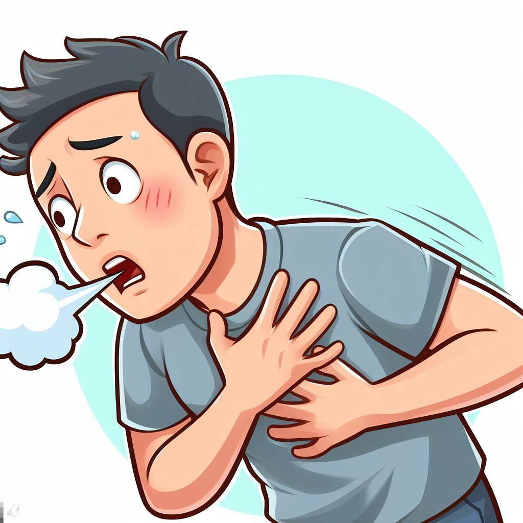 |
Imagine a bustling emergency room where a 45-year-old individual, previously robust and without significant medical history, arrives with a mysterious set of symptoms.
The patient reports a sudden onset of shortness of breath, a symptom that immediately raises concern. Upon initial assessment, vital signs paint a picture of physiological distress: a heart rate of 110 bpm, respiratory rate of 24 breaths per minute, blood pressure at 130/80 mmHg, and oxygen saturation hovering around 92% on room air. The patient appears visibly anxious, with beads of sweat forming on their forehead.
Chest auscultation reveals ominous crackles in the lower lung fields, and the patient winces with each breath.. There are no overt signs of trauma, and the lower extremities, initially, seemingly unremarkable, show no evidence of swelling or redness.
Delving into the patient’s history, we discover that they recently underwent surgery for a fractured leg. Postoperatively, prophylactic anticoagulation was prescribed. However, the patient admits to non-compliance, expressing concerns about potential bleeding risks associated with the anticoagulant therapy.
ECG Findings:
The electrocardiogram reveals sinus tachycardia with a heart rate of 110 bpm with no ST segment abnormalities
CXR Findings:
A chest X-ray exhibits a classic wedge-shaped pleural-based opacity at the periphery of the lung
What is the diagnosis?
A. Acute Respiratory Distress Syndrome (ARDS)
B. Myocardial Infarction
C. Pneumonia
D. Pulmonary Embolism
What is the pathophysiological basis for the radiographic finding?
A. Pulmonary hemorrhage
B. Alveolar consolidation
C. Infarction due to pulmonary embolism
D. Atelectasis
The most appropriate next step in the diagnostic evaluation of this patient is:
A. Ventilation-perfusion (V/Q) scan
B. Magnetic resonance imaging (MRI) of the chest
C. Transthoracic echocardiogram
D. Spiral computed tomography (CT) pulmonary angiography
Answers
1. D
2. C
Explanation:
The peripheral, wedge-shaped opacity seen on chest X-ray and confirmed by CT pulmonary angiography is indicative of infarction due to pulmonary embolism. This radiographic appearance occurs as a result of compromised blood supply to a segment of lung tissue supplied by the embolized pulmonary artery.
- Pulmonary hemorrhage may present with diffuse opacities rather than a well-defined wedge shape.
- Alveolar consolidation is characterized by homogenous opacification, not the peripheral wedge-shaped pattern seen in infarction.
- Atelectasis may present with linear or band-like opacities, but is not typically well-circumscribed with a convex border.
3. D
Given the clinical suspicion for pulmonary embolism (PE), the most appropriate initial imaging study is spiral CT pulmonary angiography.
I hope you did well and crushed this quiz.
If you need more review, click here for some free resources, questions and videos regarding pulmonary embolism
Thats all for today.
For more newsletters and case studies click here
Thanks for reading,